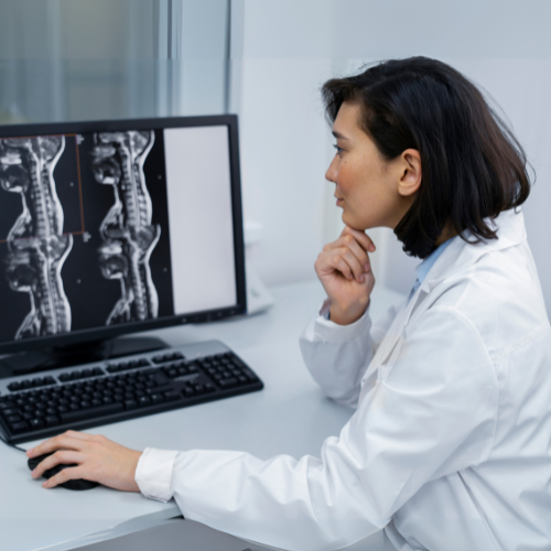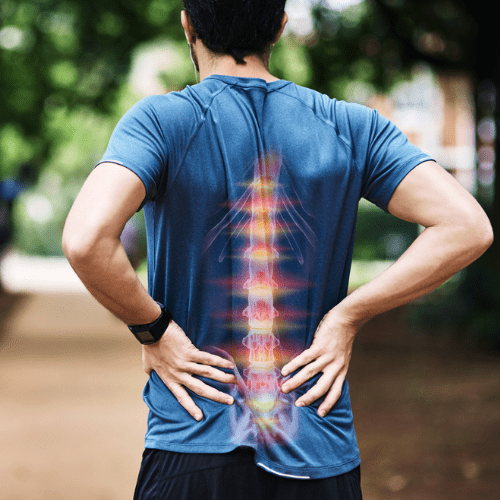
Comprehensive Care for Neck and Back Pain
Orthopedic Associates of Dutchess County leads the region in treating neck and back pain. We have the latest treatments for disc problems, nerve pain, and other spine conditions and injuries. We treat all areas of the spine:
- Cervical (neck)
- Thoracic (upper back)
- Lumbar (low back)
- Sacral (just above the tailbone) and sacroiliac (joint between spine and pelvis) pain.
- Coccyx (tailbone)
We offer nonsurgical and surgical care including therapies and minimally invasive procedures that help you move better. Back pain can interfere with every area of your life. We’re here to help you get your life back.
Why Choose Orthopedic Associates of Dutchess County for Spinal Care?
We’re known in the region as the premier practice for spine care. We get to know people personally, so that we can tailor your treatment to your goals. We offer:
- Fellowship-trained surgeons: Our spine surgeons have the highest level of training in both surgical and nonsurgical treatment. Though many people who seek care won’t need surgery, they still benefit from our spine expertise.
- Nonsurgical spine care expertise: A physiatrist or physical medicine and rehabilitation specialist works with you to maximize physical function and eliminate pain. We offer a range of nonsurgical options (including spinal injections) to help improve your neck and back pain.
- Latest advancements in treatment: We offer cutting-edge therapies and newer, less invasive surgical techniques that help you heal faster.
- Convenience every step of the way: We do everything we can to help you navigate the treatment and recovery process.
Spine Conditions We Treat
We treat both children and adults with spinal conditions such as:
- Compression fractures of the spine
- Degenerative disc disease
- Herniated or bulging disc
- Osteoporosis
- Nerve pain
- Sacroiliac joint dysfunction
- Sciatica, or radiating pain, numbness, or weakness down the front or back of the leg.
- Spinal curvature problems, such as scoliosis and kyphosis.
- Spinal stenosis, a narrowing of your spinal column.
- Spondylolisthesis, when one of your vertebrae slips out of place.
- Spondylolysis, a stress fracture of the spine.
- Spondylosis, degenerative arthritis of the spine.
Treatments for Back and Neck Pain
When we can, we start with conservative treatments, such as:
- Medications (oral or topical) to reduce pain, inflammation, or swelling.
- Injections that relieve pain, such as epidural steroid injections, facet injections and sacroiliac injections.
- Physical therapy – Working with designated physical therapy locations in the area can help reduce pain and improve mobility. For many people, PT alone can help them get back to activities they enjoy.
- Radiofrequency ablation (RFA) is a minimally invasive treatment that uses heat to disrupt nerve signals and provide relief from chronic pain.
- Alternative treatment options include bracing, chiropractic care, or acupuncture.
Spinal Surgeries We Perform
Some injuries and conditions need more intervention, including surgery. Common procedures include:
- Cervical disc replacement, also known as artificial disc replacement (ADR) or total disc replacement: A surgical procedure that replaces damaged discs in the neck with artificial discs. The goal of ADR is to restore normal disc height, relieve nerve compression, and preserve neck motion.
- Correcting spinal curvatures: Surgery is often the best option for conditions like scoliosis and kyphosis if the curve is large enough.
- Kyphoplasty: A minimally invasive procedure that treats painful compression fractures in the spine by injecting bone cement into a fractured vertebra. The procedure involves inserting a balloon into the fractured bone to create space and then filling it with cement. The goal is to relieve pain and stabilize the fracture.
- Micro Discectomy: Removing all or of part of a disc that’s bulging.
- Minimally invasive spine surgery: This type of surgery uses smaller incisions than traditional (“open”) surgery. We use this approach whenever we can because people heal faster.
- Spinal cord stimulators: We implant small devices near the spine that deliver electrical pulses to block pain signals, providing relief from chronic pain.
- Spinal decompression surgery: A surgery that reduces the pressure on the spinal cord from discs or nerves pushing against it.
- Spinal fusion: Connecting two or more bones in your spine, so that they act as a single bone.
We treat both children and adults with spinal conditions such as:
- Compression fractures of the spine
- Degenerative disc disease
- Herniated or bulging disc
- Osteoporosis
- Nerve pain
- Sacroiliac joint dysfunction
- Sciatica, or radiating pain, numbness, or weakness down the front or back of the leg.
- Spinal curvature problems, such as scoliosis and kyphosis.
- Spinal stenosis, a narrowing of your spinal column.
- Spondylolisthesis, when one of your vertebrae slips out of place.
- Spondylolysis, a stress fracture of the spine.
- Spondylosis, degenerative arthritis of the spine.
When we can, we start with conservative treatments, such as:
- Medications (oral or topical) to reduce pain, inflammation, or swelling.
- Injections that relieve pain, such as epidural steroid injections, facet injections and sacroiliac injections.
- Physical therapy – Working with designated physical therapy locations in the area can help reduce pain and improve mobility. For many people, PT alone can help them get back to activities they enjoy.
- Radiofrequency ablation (RFA) is a minimally invasive treatment that uses heat to disrupt nerve signals and provide relief from chronic pain.
- Alternative treatment options include bracing, chiropractic care, or acupuncture.
Some injuries and conditions need more intervention, including surgery. Common procedures include:
- Cervical disc replacement, also known as artificial disc replacement (ADR) or total disc replacement: A surgical procedure that replaces damaged discs in the neck with artificial discs. The goal of ADR is to restore normal disc height, relieve nerve compression, and preserve neck motion.
- Correcting spinal curvatures: Surgery is often the best option for conditions like scoliosis and kyphosis if the curve is large enough.
- Kyphoplasty: A minimally invasive procedure that treats painful compression fractures in the spine by injecting bone cement into a fractured vertebra. The procedure involves inserting a balloon into the fractured bone to create space and then filling it with cement. The goal is to relieve pain and stabilize the fracture.
- Micro Discectomy: Removing all or of part of a disc that’s bulging.
- Minimally invasive spine surgery: This type of surgery uses smaller incisions than traditional (“open”) surgery. We use this approach whenever we can because people heal faster.
- Spinal cord stimulators: We implant small devices near the spine that deliver electrical pulses to block pain signals, providing relief from chronic pain.
- Spinal decompression surgery: A surgery that reduces the pressure on the spinal cord from discs or nerves pushing against it.
- Spinal fusion: Connecting two or more bones in your spine, so that they act as a single bone.
Physicians
- Trauma and Fracture Care
- Spine
Practicing in:
Poughkeepsie and Hopewell Junction
- Spine
- Pain Management
Practicing in:
Kingston, New Windsor, Poughkeepsie, and Rhinebeck

- Pain Management
- Spine
Practicing in:
Poughkeepsie, New Windsor, and Hopewell Junction

- Pain Management
- Spine
Practicing in:
Poughkeepsie, Hopewell Junction, and New Windsor
- Spine
Practicing in:
Poughkeepsie, Hopewell Junction, New Windsor, and Kingston